国立情報学研究所 - ディジタル・シルクロード・プロジェクト
| |||||||||
|
| Explorations in Turkestan : Expedition of 1904 : vol.2 | |
| トルキスタンの調査 1904年 : vol.2 |
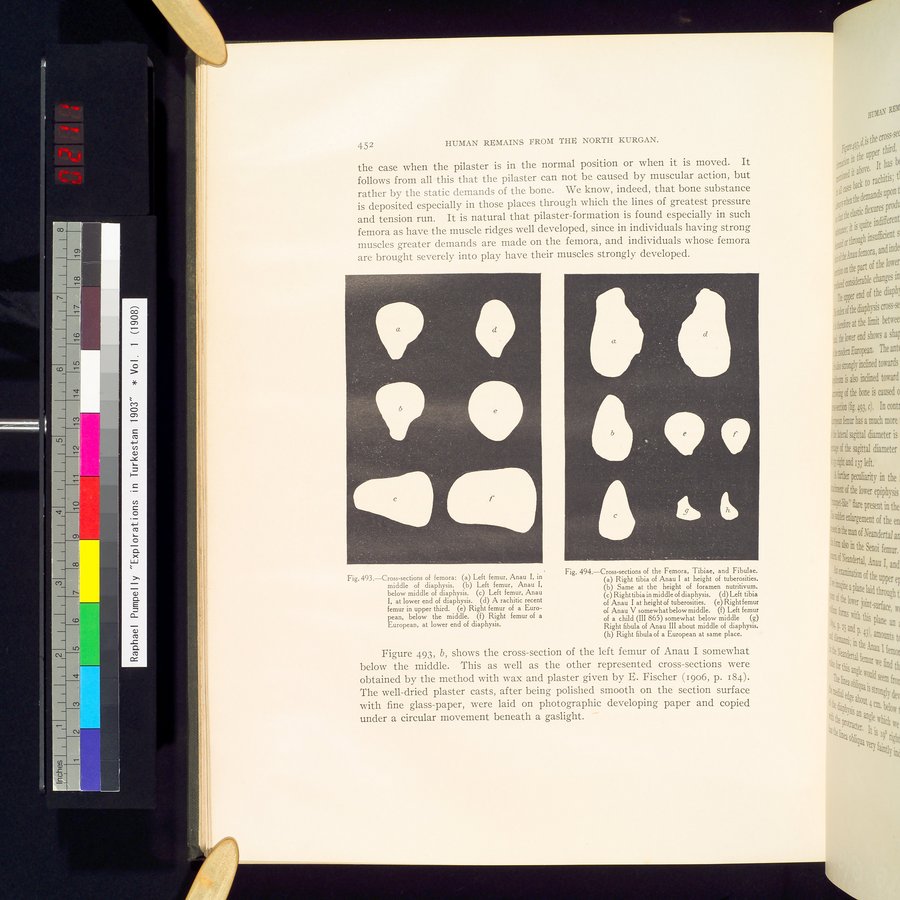 |
| 大腿骨の横断面:左大腿骨 アナウI 骨幹の中心Cross-sections of femora: Left femur, Anau I, in middle of diaphysis. | |
| 大腿骨の横断面:左大腿骨 アナウI 骨幹の下中央Cross-sections of femora: Left femur, Anau I, below middle of diaphysis. | |
| 大腿骨の横断面:左大腿骨 アナウI 骨幹の底部Cross-sections of femora: Left femur, Anau I, at lower end of diaphysis. | |
| 大腿骨の横断面:くる病にかかった最近の大腿骨上 1/3Cross-sections of femora: A rachitic recent femur in upper third. | |
| 大腿骨の横断面:欧州人の右大腿骨 下中央Cross-sections of femora: Right femur of a European, below the middle. | |
| 大腿骨の横断面:欧州人の右大腿骨 骨幹の底部Cross-sections of femora: Right femur of a European, at lower end of diaphysis. | |
| 大腿骨の横断面 脛骨と腓骨 アナウIの右脛骨粗面の最頂部Cross-sections of the femora, Tibiae, and Fibulae. Right tibia of Anau I at height of tuberosities. | |
| 大腿骨の横断面 脛骨と腓骨同じものの栄養孔の最頂部Cross-sections of the femora, Tibiae, and Fibulae. Same at the height of foramen nutritivum. | |
| 大腿骨の横断面 脛骨と腓骨右脛骨 骨幹中央Cross-sections of the femora, Tibiae, and Fibulae. Right tibia in middle of diaphysis. | |
| 大腿骨の横断面 脛骨と腓骨アナウIの左脛骨 粗面の最頂部Cross-sections of the femora, Tibiae, and Fibulae. Left tibia of Anau I at height of tuberosities. | |
| 大腿骨の横断面 脛骨と腓骨アナウVの右大腿骨 やや下中央Cross-sections of the femora, Tibiae, and Fibulae. Right femur of Anau V somewhat below middle. | |
| 大腿骨の横断面 脛骨と腓骨子供の左大腿骨(III865)やや下中央Cross-sections of the femora, Tibiae, and Fibulae. Left femur of a child (III865) somewhat below middle | |
| 大腿骨の横断面 脛骨と腓骨アナウIIIの右腓骨 骨幹のほぼ中央Cross-sections of the femora, Tibiae, and Fibulae. Right fibula of Anau III about middle of diaphysis. | |
| 大腿骨の横断面 脛骨と腓骨欧州人の右腓骨 同じ場所Cross-sections of the femora, Tibiae, and Fibulae. Right fibula of a European at same place. |
452 HUMAN REMAINS FROM THE NORTH KURGAN.
the case when the pilaster is in the normal position or when it is moved. It follows from all this that the pilaster can not be caused by muscular action, but rather by the static demands of the bone. We know, indeed, that bone substance is deposited especially in those places through which the lines of greatest pressure and tension run. It is natural that pilaster-formation is found especially in such femora as have the muscle ridges well developed, since in individuals having strong muscles greater demands are made on the femora, and individuals whose femora are brought severely into play have their muscles strongly developed.
IS
a.
d
b
a
d
b
Fig.493.—Cross-sections of femora: (a) Left femur, Anau I, in middle of diaphysis. (b) Left femur, Anau I, below middle of diaphysis. (c) Left femur, Anau
I, at lower end of diaphysis. (d) A rachitic recent femur in upper third. (e) Right femur of a European, below the middle. (f) Right femur of a European, at lower end of diaphysis.
Fig. 494.—Cross-sections of the Femora, Tibiae, and Fibulae.
Right tibia of Anau I at height of tuberosities.
Same at the height of foramen nutritivum.
Right tibia in middle of diaphysis. (d) Left tibia of Anau I at height of tuberosities. (e) Right femur of Anau V somewhat below middle. (f) Left femur of a child (III 865) somewhat below middle (g) Right fibula of Anau III about middle of diaphysis. (h) Right fibula of a European at same place.
Figure 493, b, shows the cross-section of the left femur of Anau I somewhat below the middle. This as well as the other represented cross-sections were obtained by the method with wax and plaster given by E. Fischer (1906, p. 184). The well-dried plaster casts, after being polished smooth on the section surface with fine glass-paper, were laid on photographic developing paper and copied under a circular movement beneath a gaslight.
|
Copyright (C) 2003-2019
National Institute of Informatics(国立情報学研究所)
and
The Toyo Bunko(東洋文庫). All Rights Reserved.
本ウェブサイトに掲載するデジタル文化資源の無断転載は固くお断りいたします。