国立情報学研究所 - ディジタル・シルクロード・プロジェクト
| |||||||||
|
| Explorations in Turkestan : Expedition of 1904 : vol.2 | |
| トルキスタンの調査 1904年 : vol.2 |
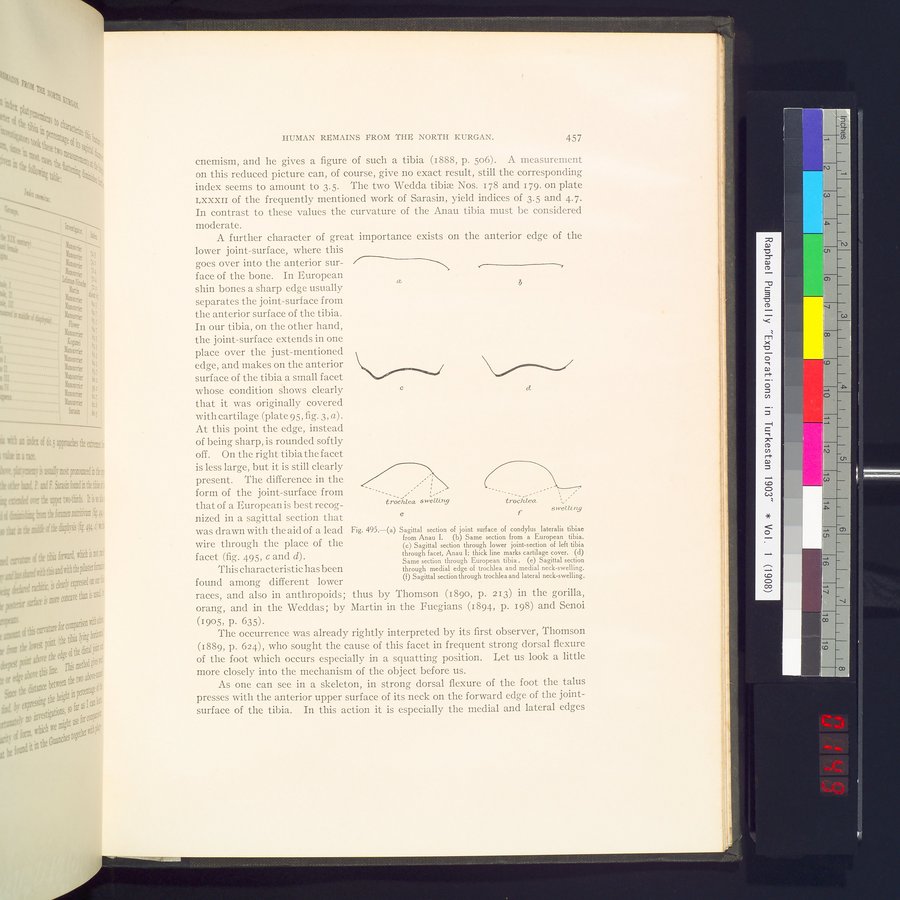 |
| アナウIで出土した外側顆脛骨の関節面の矢状部Sagittal section of joint surface of condylus lateralis tibiae from Anau I. | |
| 欧州人の脛骨の同じ部分Same section from a European tibia. | |
| 左脛骨面の低部関節面の矢状断 アナウI;軟骨膜に太線で印Sagittal section through lower joint-section of left tibia through facet, Anau I; thick line marks cartilage cover. | |
| 欧州人の脛骨の同じ断面Same section through European tibia. | |
| 内側縁滑車と内側頸部腫脹の矢状断Sagittal section through medial edge of trochlea and medial neck-swelling. | |
| 滑車と側頸部腫脹の矢状断Sagittal section through trochlea and lateral neck-swelling. |
HUMAN REMAINS FROM THE NORTH KURGAN. 457
cnemism, and he gives a figure of such a tibia (1888, p. 506). A measurement on this reduced picture can, of course, give no exact result, still the corresponding
index seems to amount to 3.5. The two Wedda tibiæ Nos. 178 and 179, on plate LXXXII of the frequently mentioned work of Sarasin, yield indices of 3.5 and 4.7. In contrast to these values the curvature of the Anau tibia must be considered moderate.
A further character of great importance exists on the anterior edge of the
lower joint-surface, where this goes over into the anterior surface of the bone. In European shin bones a sharp edge usually separates the joint-surface from the anterior surface of the tibia. In our tibia, on the other hand, the joint-surface extends in one place over the just-mentioned edge, and makes on the anterior surface of the tibia a small facet whose condition shows clearly that it was originally covered with cartilage (plate 95, fig. 3, a) . At this point the edge, instead of being sharp, is rounded softly off. On the right tibia the facet is less large, but it is still clearly present. The difference in the
form of the joint-surface from
that of a European is best recog- trochlea swelun9
nized in a sagittal section that
was drawn with the aid of a lead Fig. 495.—(a) Sagittal section of joint surface of condylus lateralis tibiae
from Anau I. (b) Same section from a European tibia.
wire through the place of the
(c) Sagittal section through lower joint-section of left tibia
facet (fig. 495, c and d). through facet, Anau I; thick line marks cartilage cover. (d)
Same section through European tibia . (e) Sagittal section
This characteristic has been through medial edge of trochlea and medial neck-swelling.
(f) Sagittal section through trochlea and lateral neck-swelling.
found among different lower races, and also in anthropoids; thus by Thomson (189o, p. 213) in the gorilla, orang, and in the Weddas; by Martin in. the Fuegians (1894, p. 198) and Senoi
(1905, p. 635).
The occurrence was already rightly interpreted by its first observer, Thomson (1889, p. 624), who sought the cause of this facet in frequent strong dorsal flexure of the foot which occurs especially in a squatting position. Let us look a little more closely into the mechanism of the object before us.
As one can see in a skeleton, in strong dorsal flexure of the foot the talus presses with the anterior upper surface of its neck on the forward edge of the joint-surface of the tibia. In this action it is especially the medial and lateral edges
y
b
C
ei
e
|
Copyright (C) 2003-2019
National Institute of Informatics(国立情報学研究所)
and
The Toyo Bunko(東洋文庫). All Rights Reserved.
本ウェブサイトに掲載するデジタル文化資源の無断転載は固くお断りいたします。