National Institute of Informatics - Digital Silk Road Project
| |||||||||
|
| Explorations in Turkestan : Expedition of 1904 : vol.2 |
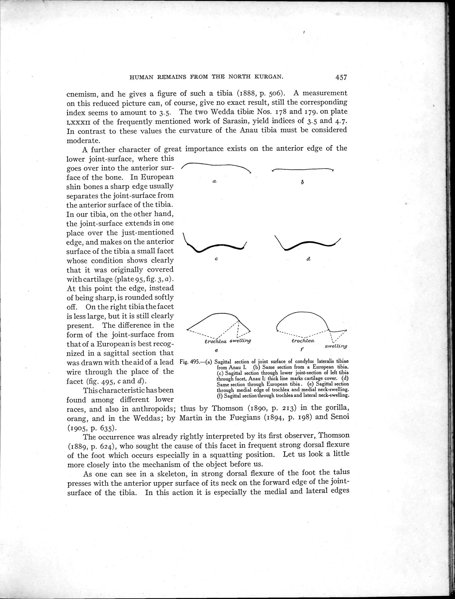 |
| Sagittal section of joint surface of condylus lateralis tibiae from Anau I. | |
| Same section from a European tibia. | |
| Sagittal section through lower joint-section of left tibia through facet, Anau I; thick line marks cartilage cover. | |
| Same section through European tibia. | |
| Sagittal section through medial edge of trochlea and medial neck-swelling. | |
| Sagittal section through trochlea and lateral neck-swelling. |
HUMAN REMAINS FROM THE NORTH KURGAN. 457
cnemism, and he gives a figure of such a tibia (1888, p. 506). A measurement on this reduced picture can, of course, give no exact result, still the corresponding
index seems to amount to 3.5. The two Wedda tibiæ Nos. 178 and 179, on plate LXXXII of the frequently mentioned work of Sarasin, yield indices of 3.5 and 4.7. In contrast to these values the curvature of the Anau tibia must be considered moderate.
A further character of great importance exists on the anterior edge of the
lower joint-surface, where this goes over into the anterior surface of the bone. In European shin bones a sharp edge usually separates the joint-surface from the anterior surface of the tibia. In our tibia, on the other hand, the joint-surface extends in one place over the just-mentioned edge, and makes on the anterior surface of the tibia a small facet whose condition shows clearly that it was originally covered with cartilage (plate 95, fig. 3, a) . At this point the edge, instead of being sharp, is rounded softly off. On the right tibia the facet is less large, but it is still clearly present. The difference in the
form of the joint-surface from
that of a European is best recog- trochlea swelun9
nized in a sagittal section that
was drawn with the aid of a lead Fig. 495.—(a) Sagittal section of joint surface of condylus lateralis tibiae
from Anau I. (b) Same section from a European tibia.
wire through the place of the
(c) Sagittal section through lower joint-section of left tibia
facet (fig. 495, c and d). through facet, Anau I; thick line marks cartilage cover. (d)
Same section through European tibia . (e) Sagittal section
This characteristic has been through medial edge of trochlea and medial neck-swelling.
(f) Sagittal section through trochlea and lateral neck-swelling.
found among different lower races, and also in anthropoids; thus by Thomson (189o, p. 213) in the gorilla, orang, and in the Weddas; by Martin in. the Fuegians (1894, p. 198) and Senoi
(1905, p. 635).
The occurrence was already rightly interpreted by its first observer, Thomson (1889, p. 624), who sought the cause of this facet in frequent strong dorsal flexure of the foot which occurs especially in a squatting position. Let us look a little more closely into the mechanism of the object before us.
As one can see in a skeleton, in strong dorsal flexure of the foot the talus presses with the anterior upper surface of its neck on the forward edge of the joint-surface of the tibia. In this action it is especially the medial and lateral edges
y
b
C
ei
e
|
Copyright (C) 2003-2019 National Institute of Informatics and The Toyo Bunko. All Rights Reserved.