National Institute of Informatics - Digital Silk Road Project
| |||||||||
|
| Explorations in Turkestan : Expedition of 1904 : vol.2 |
 |
| Cross-sections of femora: Left femur, Anau I, in middle of diaphysis. | |
| Cross-sections of femora: Left femur, Anau I, below middle of diaphysis. | |
| Cross-sections of femora: Left femur, Anau I, at lower end of diaphysis. | |
| Cross-sections of femora: A rachitic recent femur in upper third. | |
| Cross-sections of femora: Right femur of a European, below the middle. | |
| Cross-sections of femora: Right femur of a European, at lower end of diaphysis. | |
| Cross-sections of the femora, Tibiae, and Fibulae. Right tibia of Anau I at height of tuberosities. | |
| Cross-sections of the femora, Tibiae, and Fibulae. Same at the height of foramen nutritivum. | |
| Cross-sections of the femora, Tibiae, and Fibulae. Right tibia in middle of diaphysis. | |
| Cross-sections of the femora, Tibiae, and Fibulae. Left tibia of Anau I at height of tuberosities. | |
| Cross-sections of the femora, Tibiae, and Fibulae. Right femur of Anau V somewhat below middle. | |
| Cross-sections of the femora, Tibiae, and Fibulae. Left femur of a child (III865) somewhat below middle | |
| Cross-sections of the femora, Tibiae, and Fibulae. Right fibula of Anau III about middle of diaphysis. | |
| Cross-sections of the femora, Tibiae, and Fibulae. Right fibula of a European at same place. |
452 HUMAN REMAINS FROM THE NORTH KURGAN.
the case when the pilaster is in the normal position or when it is moved. It follows from all this that the pilaster can not be caused by muscular action, but rather by the static demands of the bone. We know, indeed, that bone substance is deposited especially in those places through which the lines of greatest pressure and tension run. It is natural that pilaster-formation is found especially in such femora as have the muscle ridges well developed, since in individuals having strong muscles greater demands are made on the femora, and individuals whose femora are brought severely into play have their muscles strongly developed.
IS
a.
d
b
a
d
b
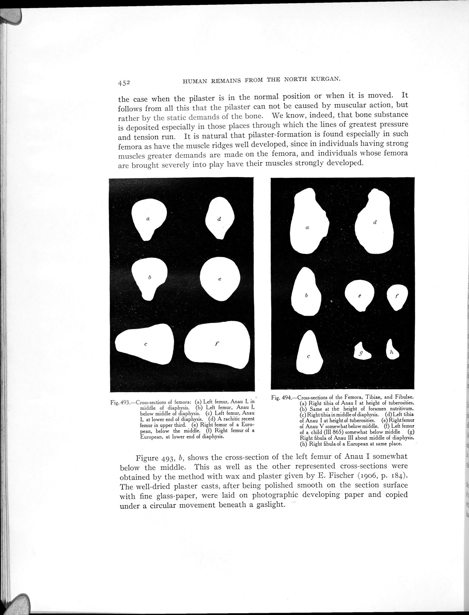
Fig.493.—Cross-sections of femora: (a) Left femur, Anau I, in middle of diaphysis. (b) Left femur, Anau I, below middle of diaphysis. (c) Left femur, Anau
I, at lower end of diaphysis. (d) A rachitic recent femur in upper third. (e) Right femur of a European, below the middle. (f) Right femur of a European, at lower end of diaphysis.
Fig. 494.—Cross-sections of the Femora, Tibiae, and Fibulae.
Right tibia of Anau I at height of tuberosities.
Same at the height of foramen nutritivum.
Right tibia in middle of diaphysis. (d) Left tibia of Anau I at height of tuberosities. (e) Right femur of Anau V somewhat below middle. (f) Left femur of a child (III 865) somewhat below middle (g) Right fibula of Anau III about middle of diaphysis. (h) Right fibula of a European at same place.
Figure 493, b, shows the cross-section of the left femur of Anau I somewhat below the middle. This as well as the other represented cross-sections were obtained by the method with wax and plaster given by E. Fischer (1906, p. 184). The well-dried plaster casts, after being polished smooth on the section surface with fine glass-paper, were laid on photographic developing paper and copied under a circular movement beneath a gaslight.
|
Copyright (C) 2003-2019 National Institute of Informatics and The Toyo Bunko. All Rights Reserved.